
Dr.
Niamtu
|
Rancho Mirage, Calif. - Failure to
address the midface when performing facial rejuvenation can
lead to imbalance and patient dissatisfaction, said
oral/maxillofacial and cosmetic facial surgeon Joseph Niamtu
at the Annual Scientific Meeting of the American Academy of
Cosmetic Surgery.
He described his own
simplified technique for minimally invasive midface lift with
optional simultaneous augmentation of the nasolabial folds,
stressing that when performing facial rejuvenation, at any
level, it is important to consider what you're doing in the
context of the entire face.
Dr. Niamtu said that the
midface-lifting technique that he currently uses requires
minimal incisions, which are hidden. This procedure causes no
negative effect on lower eyelid position, and can actually
optimize conditions for a problematic lower lid, he said,
adding that the procedure is easily performed in an ambulatory
environment.
Specifically, it requires
incisions both intraorally as well as in the temporal tuft.
With experience, he said the procedure can eventually be
performed in approximately 15 minutes per side.
The initial 2-cm temporal
incision, located in a similar location to that used for an
endoscopic brow lift, is first marked and then completed in
the temporal tuft, perpendicular to the alar-canthal line,
down to the level of the superficial layer of the deep
temporal fascia.
Once the temporal incision
is made, a dissection is performed with a No. 9 periosteal
elevator in a line that intersects the lateral orbital rim and
frontozygomatic suture.
When the lateral orbital
rim is encountered, the elevator is rotated and used to burrow
under the periosteal layer. This dissection plane proceeds
from the deep layer of the superficial temporal fascia to the
subperiosteal layer, thereby avoiding damage to the frontal
branch of the facial nerve, as these are technically both
"safe" dissection planes.
After entering the
subperiosteal layer, the dissection is directed from above,
over the malar eminence, completing the temporal dissection.
Attention is then directed to the mouth. A gingival vestibular
incision is made 5 mm above the attached gingiva, in the area
of the cuspid tooth, to the first molar. Subperiosteal
dissection is then performed to the inferior orbital rim,
lateral nasal rim, and over the zygoma to the junction of the
zygomatic arch.
"Caution is used here to
avoid damage to the infraorbital nerve," he said. "This
dissection joins the pocket created by the previous temporal
dissection, thus creating a tunnel from the temple to the oral
cavity."
The malar fat pad is
readily visible and is sandwiched between the periosteum,
muscle, and oral mucosa. Once identified, it is secured with a
double-throw from a 2-0 PDS suture that will be passed up
through the dissection tunnel.
"The position where the
malar fat pad is secured with this suture relates to the final
vector of the lift," Dr. Niamtu said. "By securing the fat pad
in the region of the canine tooth, a more superior lift is
produced. However, securing the fat pad in the region of the
first molar produces a more superior lateral augmentation."
According to Dr. Niamtu,
the ability to adjust the vector of the mid-facial
augmentation is the main advantage of this technique. He said
this allows variability and customization of the cosmetic
result and is the perfect midface "implant."
Dr. Niamtu generally
recommends securing the fat pad in the first molar area. After
the suture is threaded, the position can be redirected on the
fat pad to adjust to the desired level of augmentation.
"In some instances I will
use two sutures: one in the cuspid region and one in the first
molar region for a maximum augmentation," he said.
The next step is to pass
the suture back through the incision tunnel. A thin tonsil
clamp or a passing awl is positioned and passed from the
temporal incision to the intra-oral area. The needle is cut
off the suture and both suture tails are secured with the
clamp or awl. The suture tails are then pulled back through
the temporal incision, placed under tension, and the elevation
of the midface is immediately apparent.
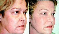
Patient pre-op (left) and post-op
(right) after undergoing a simplified mid-face lifting
technique that requires minimal incisions, which are
hidden.
|
"The more tension placed on the
suture tails, the more the midface is elevated," he said. "The
level of augmentation may appear excessive, as the patient is
in a recumbent position; however, post-operatively this is
rarely the case."
Attachments designed to
last
Dr. Niamtu secures the
sutures with maximum fat pad elevation. Once he is satisfied
with the vector of the lift, Dr. Niamtu threads a passing
needle on the suture end and secures it to the superficial
layer of the deep temporalis fascia, now under tension, to
maintain the elevated midface. When the surgical site heals,
the repositioned periosteum and soft tissues will reattach for
a lasting augmentation.
In consideration of further
mid-facial rejuvenation, Dr. Niamtu frequently addresses the
nasolabial folds with Gore-Tex implants or fat transfer, and,
in certain cases CO2 laser resurfacing.
"There is one caveat," Dr.
Niamtu said. "Due to the extreme amount of elevation achieved,
sometimes the intra-oral incision is gaping under the tension,
with seemingly insufficient tissue to close the incision. If
this should occur, simply dissecting the tethered mucosa from
the deeper tissue will enable primary closure of the wound."
Dr. Niamtu said that if
concomitant lower eyelid surgery is planned, the midface lift
should be performed first because elevating the midface can
change the lower eyelid esthetics.
Due to subperiosteal
dissection, swelling can persist from one to three weeks. Most
patients will experience a temporary paresthesia in the
distribution of the infraorbital nerve. Dr. Niamtu said that
he has not seen permanent numbness from this procedure, as
this nerve can be easily seen and avoided during
surgery.
"Because of the release of
the periosteum, and subsequently the origin of the lip
elevators, a temporary dysfunction may be seen upon smiling or
puckering," he said. "In my experience, this improves over a
several week period. This doesn't usually present as a problem
if the patient is properly forewarned."
Dr. Niamtu said that he has
seen two cases of intraoral wound dehiscence, both in smokers.
This was treated by wound hygiene, using peroxide and
antibiotic oral rinses. In both cases healing was uneventful.
He said that another
possible option for midface rejuvenation involves the use of
facial implants, but some patients are uncomfortable with
thought of a foreign substance in their face.
"The minimally invasive
rejuvenation procedure is easier to accept for some patients
than the use of facial implants," he said. "The procedure
consists of a repositioning of the patient's own tissue by
elevating the malar fat pad. This procedure is not
appropriate, however, for individuals who have a very thin,
wasted or atrophic midface. In these cases there's no fat pad
to lift. This is not a procedure for patients who are very
gaunt, without adequate midface tissue."
Dr. Niamtu is currently
doing a study on a cohort of 20 consecutive midface lift
patients and, as this group approaches the two-year
postoperative point, he said the level of augmentation and
patient and operator satisfaction remain high.
